clovek

MY ACCOUNT

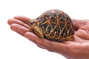
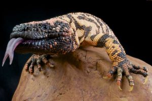
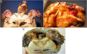
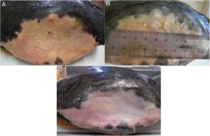
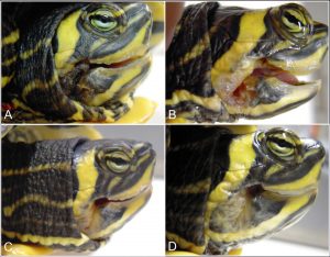
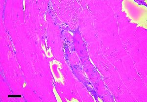
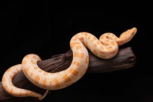
Here is the worm inside


reptiliendoktor-245166985wurm1

Almost all wild animals are infected with parasites. Under natural conditions, this is not a problem, because the host and parasite have adapted themselves over many millennia. A natural balance has been established between them.

Artificial living conditions in the terrarium or freights, however, alter this balance in favor of the parasites. Spatial limitation and close socialization mean that the animals are in constant contact with the eggs, spores or cysts of the parasites. The high excitation pressure can lead to mass infestation. A harmless parasite will quickly become a health threat.

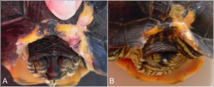
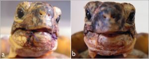
The parasite attack can vary greatly depending on the conditions and the stress. Sometimes it is continually enhanced by continual new infection, but it may also suddenly increase explosively, e.g. When additional stresses such as stress or disease are added. Typical symptoms are:

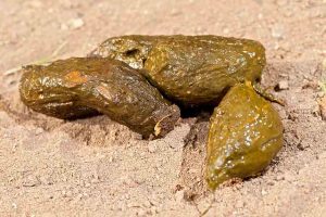
Digestive disturbances, recognizable by soft, pulpy or liquid faeces. Growth disturbances in young animals
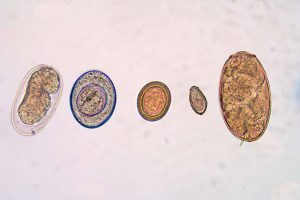
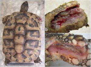
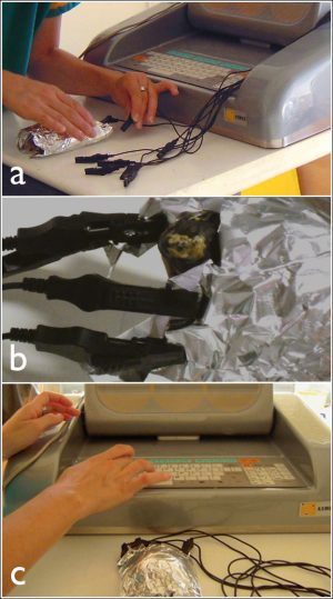
 

reptiliendoktor-298524680wurm2

Fecal examination

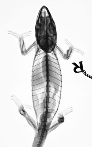
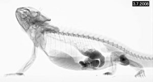
Regular faecal examinations are one of the most important medical measures in terrarium management. Since parasites and their eggs are generally excreted in droppings, pathogenic pathogens can be reliably identified in this manner and specifically controlled by specific drugs. As a rule, it is sufficient to examine outwardly healthy animals once a year, in the case of chronic infestations or increased exciters, twice a year. Species holding a winter or summer rest are examined no later than two months before the start of the rest period.

Wild catches should definitely be investigated immediately after purchase, even if the animals seem to be healthy. Until the diagnosis or successful treatment they are In order not to introduce any diseases into the old stock.

Treatment

The therapy should always be carried out by a reptile veterinarian, since preparation and dosage can differ from the regulations for mammals. Unfortunately, many of the agents used kill not only pathogenic single cells, but also useful intestinal symbionts and thus lead to damage to the intestinal flora. Accompanying the drug treatment should therefore always be respected.

Affected animals are separated and placed in a minimally set-up area during the entire treatment period to pay attention. To avoid re-infections, all excretory products must be removed quickly from the pelvis.

In most cases terrarium, soil substrate and equipment must be thoroughly disinfected and the terrarium plants must be disposed of.

reptiliendoktor-232568680wurm3

www.reptiliendoktor.com

Translation Heike Krüger, edit www.terraristik.cz

NEW OPTIONS FOR THE PRESENTATION

We offer you the possibility of having your animals, company, your product or e.g. to present a trade fair or stock exchange. In the case of a stock exchange, you are more likely to reach potential visitors. For more attention, you can offer footage from previous events. Interested people like to look at the place, the environment and of course the offered animals. All dynamic, as video not only the simple pictures. Similarly, the photo galleries of your animals that you offer for sale will attract more attention when offered in the form of a mix of subtitled photos. You can discuss the video with their words or accompany them with background music. (Beware of a potential copyright conflict! Youtube.com can check the level of protection, various titles). We offer you the option to create a video guaranteeing copyright for the selected background music. Details will be provided upon request (info@terraristik.cz) You must register for the offer of advertising and use other services and options on our website. You can insert videos in the Links section, then add the link and select the link. (The option Standard is the publication of the base link in the left column is free). The link Exclusiv, which also publishes your ad in the field of ads (banner column), but also in the areas of reptile club and photo gallery. Please enter a form for entering lines to open the link to edit the text. When you insert a video, select the Video Selection check box. This option adds a video icon to your image, which makes it clear that it is not a “simple” link to a website. Links can be firmly positioned like advertisements and so they have the most advantageous positions. Everything for a symbolic price of  1 € / 3 day. You have the option of editing, expanding and deleting all lines. If any lines containing racist, pornographic or other unwanted content appear, you can retrieve them immediately and without refund. We also enjoy the classic banner (link to web pages) as well as banner with a link to a video (banner will be a video icon) In our case, a short description or a banner title is included. Further information on request.

Examples of a possible presentation

Robert Schlesinger, admin Terraristik.cz


Diseases in reptiles


reptiliendoktor-241196731-1

Reptiles show a disease first in behavioral changes: often the affected animal reduces the food intake or no longer eats at all. In addition, one can observe changes in the activity: the animal moves less or avoids certain movements. Sometimes it sleeps a lot or withdraws completely.

In addition, each species reacts in a manner that is characteristic for them: many lizards and snakes become more aggressive. In chameleons the skin color changes. Aquatic turtles lie with their eyes closed in the vicinity of the heat lamp, are less likely to enter the water or have problems with swimming and diving. Land turtles tend to bury themselves in disease. In snakes, symptoms such as moles or moistening problems can point to existing diseases.

In addition to these general symptoms, there are a large number of specific disease symptoms. These indicate the path to certain diseases. You will find all the information in our Symptom-Finder.

reptiliendoktor-178344272-2

More than 90% of all reptile diseases are caused by suboptimal living conditions – this shows how important a proper animal welfare is for the health of our reptiles.

If an animal is already ill, usually only the veterinarian can help. In addition, however, a consequent improvement in posture conditions is indispensable – otherwise the disease can break out again.

The effort is rewarded with certainty because even minor improvements in posture can produce great effects.


Diseases of Greek land turtle and other land turtle species

reptiliendoktor-118292470-3

Common: kidney disease, gout, overweight, dehydration, rickets

Uncommon: liver disease, diarrhea, constipation, respiratory disease, coryza, parrot, urinary tract, urinary tract infections, bot skin infections, abscess, injury of the armor, skin and connective tissue injury, skin mycosis, armor infections, intestinel parasites, cloaca disease, joint disease, eye disease , Postchibernale sepsis, stomatitis, acute stress, food deficiency, emaciation, fatigue syndrome, herpes, URTD, irido virus, vitamin A overdose, vitamin D overdose, neoplasia

Typical postural deficiencies: over-supply with fat and protein, terrarium keeping, too dry attitude

Diseases of ornamental turtles, swamp turtles and other species of turtles

reptiliendoktor-393566221-4

Common: liver disease, overweight, skin and connective tissue injury, abscess, armor infection, fetal

Uncommon: Bacterial skin infections, skin mycosis, diarrhea, respiratory disease, runny nose, oestrus, vitamin A deficiency, dyspneasia, ocular disease, rickets, constipation, injury to the armor, acute stressfulness, renal disease, urinary tract infection, joint disease, cloaca disease, stomatitis, gas bladder disease , Papillomatosis, nourishment, emaciation, fatigue syndrome, neoplasia, moistening problems, vitamin B1 deficiency, vitamin D overdose, vitamin E deficiency

Typical postural abnormalities: lack of warming, overfeeding, insufficient plant food, lack of soil (female).


Diseases in cornnatter, python, boa and other snake species

reptiliendoktor-240333853-5

Common: Cropping problems, mites, bacterial skin infections, skin mycosis, stomatitis

Uncommon: Dehydration, Darmparasites, Abscess, Gastroenteritis, Eye disease, Respiratory system disease, Liver disease, Kidney disease, Constipation, Diarrhea, Amoebia, Cryptosporidia, Skin and connective tissue injury, Burns, Acute stress, Joint disease, Spondylosis, Cloccal disease, (IBD), URTD, vitamin B1 deficiency, vitamin D overdose, vitamin C deficiency, vitamin E deficiency, bladder disease, gas bladder disease

Typical postural disorders: Unhygienic posture, too dry

Illnesses in Bartagame, Leopard Gecko, Iguana, Chameleon, Anolis, Skink, Lizard, Teju and Waran

reptiliendoktor-122625310-6


Common: liver disease, overweight, skin and connective tissue injury, wound infection, abscess

Uncommon: dehydration, kidney disease, gout, constipation, acute stress, dyspnoea, coccidia, cryptosporidia, bacterial skin infections, skin mycosis, urinary tract infection, stomatitis, respiratory disease, coryza, Rashitis, joint disease, spondylosis, papillomatosis, legend, food depletion, emaciation, fatigue syndrome, neoplasia, vitamin A overdose, vitamin D overdose, vitamin B1 deficiency, vitamin C deficiency, URTD

Typical postural deficiencies: overfeeding, too dry, too close socialization

http://www.reptiliendoktor.com/

Translation Heike Krüger, edit www.terraristik.cz


FOREWORD VET

Buy a terrarium animal does not mean any problem at the present time, unfortunately. Why unfortunately? The answer is obvious. The fact that today you can easily purchase amphibian, reptiles, bird or small mammal via the Internet, on the animals exchanges, in brick-and-mortar stores or elsewhere, leading to the fact that it can take almost anyone. Thus, even a person with no previous experience and any information or at least the basic biological and zoological awareness. Remember that, for example, keeping reptiles is very challenging, and this applies to small turtles, chameleons … through to large snakes or lizards. For example, as many popular small tortoises, whether land or water (and not only those) are for their cuteness very often recommended as a “gift for children,” even though they and even their own parents do not have a clue about the complexity of such a breed.
The basic thing which should every breeder realize and understand is the fact that possession and stay in captivity the animals have not chosen voluntarily. Therefore, it is necessary to act accordingly and to provide a possible optimal and most natural conditions for their life.
In our field, we meet often tragic destinies of captive bred reptiles, and not only because of the above-mentioned demands of breeding, but mainly from the lack of obtained information about the breed of the animal before buying it. We can see animals often in such condition when for their salvation is already too late. If we manage to save the animal’s life, then it is often affected by such effects, it would not survive in the wild. It is a cruel tax for breeder´s irresponsibility, do not you think?
Basal ignorance and the consequent unwillingness to inform buyers of the animal, we meet already in the first line – so even by some retailers. Some of them actually prefer business to the interests of the animal. It is, however, up to the future breeder to get all relevant and necessary information for animal breeding himself. So should keep each responsible person who plans to take his new home ‘pet’. It cannot already make excuses use the lack of literature in printed or electronic form nowadays. Our department is not indifferent to such breeders and we are able alternatively to advise, recommend the best source of information.
If you are considering the selection and purchase of a new family member, take the following words of wisdom to heart:
“You become forever responsible for what you have bound to yourself.” (Antoine de Saint-Exupéry).

Autor: MVDr. Ing. Eva Čermáková

Principles for the nutrition of reptiles in the Pet care


It is true that reptiles have a slower metabolism than e.g. Birds or mammals, which should also be considered in the diet. Small carnivorous reptiles, also need their food adapted to their size. They get smaller portions or fodder animals and have to be fed more often. Large carnivorous reptiles, also adapted to their size their feed or feed animals. They are then fed at a greater distance and not quite as frequently as the smaller species, or young animals. Insect-eating lizards, must be fed daily. Babies have no rationed feeding, they should eat so much until they are full. Some Adult reptiles are fed 1 or 2 times a week. (Genus Elaphe, Vipera, Lampropeltis, Nerodia). Larger species are fed only 2 or 3 times a month, depending on the size of the fodder, e.g. (Boa, Pythons, Eunectes, Corallus).                                                            *


Replacement food- or masking

Some reptiles orient themselves in hunting, on the smell of their prey. In order to get young animals to feed, small mice are rubbed with fish or one rubs these babymuses over the skin of frogs. In addition, one can use the skin of lizards or snakes to prepare mice with it to give them a different smell. Mice, especially lizards or snakes, are much better tolerated by snake-eatings or by  lizard-eatings, and the conversion to a substitute food is easier. The use of protected species e.g. Amphibians or other reptiles as feed animals for exotic species is completely unacceptable.                                                        


Crocodiles

Young crocodiles can also be fed with inverted feed animals, as well as cockroaches, beetles, grasshoppers, cockchafer. Or you can take rodents, chickens or fish. Adult crocodiles are fed with rodents, chickens and fish. The risk of obesity requires high attention. The reptiles are kept far too often, in enclosed spaces with minimal active movement, which often leads to the animals becoming overweight. Therefore, the diet should be adjusted to about 8 to 10% of the body weight per week. Fish has all the necessary nutrients, including minerals and vitamins. If, at times, rodents and chickens are still fed, a deficiency of vitamin B1 is also prevented. Precautionary, if only fish is fed, can also be administered between thiamine (B1).


Turtles

Water turtles (aquatic turtles) are best fed with small creatures, insects and worms, depending on their age. Later, fish and small rodents are added and then gradually increased. It is important to always provide fresh food, which should also be absorbed quickly. Small pieces are best served. The Aquaterrarium should be divided so that you have a part where you can feed seperat, so you can pay attention to the cleanliness, the water. This method is also suitable for older animals. Water plants and snails also belong to the feed. Water plants and snails introduced by nature can lead to infection and should therefore be omitted. If yunnge turtles learn not to go to the water plants, they can get used to lettuce, cabbage, carrots or leaves. For hygienic reasons, only as much food is offered as is immediately eaten.


Land turtles (terrestrial turtles) are mostly omnivore (Omnivores)

Therefore they get fruits and vegetables, which is supplemented with animal protein (especially in young animals). The food should be provided so that it can not be contaminated with feces or urine. As a dietary supplement, calcium should be added to the diet (whether with a solution or powdered).


Replacement food for turtles

There are different types of grained and preserved feed (Mainly for turtles and iguanas). These reptiles can be accustomed gradually, but it should only be understood as a dietary supplement. It is not a complete substitute for natural fresh feed and should not be used as a main feed. From the point of view of veterinary practice, turtles are good patients, not so shy and also willing to take the necessary medication without problems.


Snakes

The basic food of the snakes in private households / breeders consist of rodents and chickens. Through the food , the snake takes up the entire body and gets everything it needs. A snake therefore does not need any additional preparations such as vitamins or minerals. Many snakes are selective about the hunted animal, and what they eat and what is not, although no significant difference can be seen in size or color, but all these animals do not eat. It is not only the smell, also the sight plays a significant role in hunting. Tests have shown that the snakes attack their prey in the terrarium, separated by a glass wall, specifically. The animals lack the naturalness and the necessity of complex hunting. The risk of obesity is great and can also involve great problems, e.g. Reduced reproductive activity, deterioration of the immune system. They may also suffer from liver and circulatory disorders. Some authors report that the feeding of reptiles with vertebrate animals occurs in a manner that is inconsistent with the principle of animal welfare and its elimination in this context with the legal restrictions. Dead beasts would reduce the risk of injury to reptiles. If some snakes accept freshly killed food, there is no reason not to use this method. There are arguments against the proposition and also efforts based on the natural needs of the reptiles:

* Many reptiles do not eat their offered food because the visual  Perception and reception, of specific smell signals

   Irreplaceable role.


* Pray fishing is an alternative, for natural reptile activity.   It is not yet known what role the individual phases of hunting in the

   Preparation for digestion (neuroendocrine regulation).


Frozen food for snakes

Preserved killed rodents and birds in a frozen state, has many disadvantages. On the one hand, the nutritional value decreases and the risk of food contamination is increased. Frostfutter, can only be an emergency solution if fresh food is not available.


Wild living vertebrates as feed animals for snakes

The reasons which lead us to immediately reject this practice are the hygienic reasons. The danger of an infection would be much too great.


Lizards carnivores

Larger species of lizards e.g. The Varan, are fed with rodents or chickens.


Lizards insectivor

Worms, centipedes, spiders, butterflies, beetles and grasshoppers are very tasty for lizards. In the summer there is therefore the best and the best food. Rice beetles, cockroaches, flies, etc. can be breeded all year round. A good calcium ratio of the phosphorus in the diet is important and should provide a good ratio of 1.5: 1 to 2: 1 in the blood of healthy reptiles. The most prevalent claims are the eggs of the pregnant females. The dose of the feed of insectivorous lizards must be supplemented with minerals. Without this measure the lizards would suffer from calcium deficiency and develop serious metabolic disorders. The situation is complicated by the lack of active vitamin D (D3). The unfavorable situation can be treated by indirect nutritional improvement, e.g. By feeding invertebrate animals, or by adding Reptiles Directly. For some groups such as geckos, it is recommended to give the feed insects with mineral supplements at each feeding. Some species, including smaller lizards (skink, chamelion, iguana, geckos, etc.) also take small rodents. But, too much of this food can cause liver and kidney problems.


Herbivore and Omnivore

Terristic species of turtles and the green iguanas (Iguana Iguana) are the most common patients with mineral metabolic disorders. The reason for this is the one-sided feeding of inappropriate food. In the diet of these reptiles we attach importance to calcium and trace elements. Iguanas like to eat flowers, fruits and juicy leaves. It is always advisable that the feed offered is varied. (Strawberries, raspberries, tomatoes, peppers, melons, apricots, peaches, grapes, bananas, hibiscus, meadows). Animal proteins are obtained by boiled eggs, insects and intestines of rodents. Adult Iguanas should not be received by the young rodents more than once a month, taking into account their movement and propagation cycles. The young animals are fed in the attitude ad libitum (they can eat until they are fed up), adults three times a week. There is special food for the genus Brachylophus, Conoluphus, Ctenosaurus, Cyclura, Dipsosaurus, Iguana and Sauromalus.


Liquid

The fluid requirements vary according to the type of fluid used (Environmental temperature, animal activity) and water content in the diet. Turkeys fed apples, tomatoes and fresh cabbage drink much less Water than when feeding oatmeal, granules and hay. The water has to be fresh and clean and also in clean containers. It should not be too cold either. The best is to take a slightly stagnant water.

Higher chlorine content can be removed by holding the water in the tank (it can also be ventilated). The gift of drugs and Supplements in drinking water can adversely affect the taste of water (vitamin A). Reptiles receive water mainly through the feed. Reptiles that feed on juicy fruits, fruits and vegetables hardly need drinking water. Snakes, turtles and some lizards drink from bowls, Small lizards take the water by taking the dew clouds and the walls Of the terrarium. The drinking bowls shall be shaped in such a way that: The water in the bowl is easily accessible (to make life-threatening Drowning). In the genus Phrynosoma the water intake between the scales on the back was repeatedly observed, from where it flows through the small canals in the skin and on the mouths. Higher demands on the fluid balance have sick reptiles, especially dehydrated ones. Patients with severe respiratory or intestinal infections. We offer this patient at least 25 ml / kg daily. In addition to the pen feed, fluids can be supplied via a sheath. An effective form of fluid balancing is also a regular bath.

Zdeněk Knotek  Prof. MVDr., CSc., Dipl. ECZM

Translation Heike Krüger,  graphic adaptation Terraristik.cz


Physical reparative treatment in reptiles

  • Salvatore Rinaldi,
  • Maddalena Iannaccone,
  • Gian Enrico Magi,
  • Emanuela Costantini,
  • Alessandro Castagna,
  • Eraldo Sanna Passino,
  • Margherita Maioli and
  • Vania Fontani

Received: 2 January 2013

Accepted: 11 February 2013

Published: 26 February 2013


Abstract

Background

The tissue growth necessary to achieve a complete or partial restitution ad integrum as a result of injury to soft tissue and/or hard times in reptiles is variable and often needs long time in relation to the species, to the habitat and to their intrinsic physiological characteristics. The purpose of this work was to see if the tissue optimization (TO) treatment with radio electric asymmetric conveyer (REAC) provided good results in these animals and whether its use translates into reduced time of tissue repair. This paper describes preliminary results with in promoting the tissue repair in reptiles.

Cases presentation

A 5 year old male Testudo graeca (Leo) and Trachemys scripta scripta (Mir) and a 15 year old female Testudo hermanni (Juta) were evaluated because of soft tissue injuries. A female 25 year old Trachemys scripta elegans (Ice), a female 2.5 year old Trachemys scripta scripta (Penelope) as well as a 50 year old male Testudo graeca (Margherito) were evaluated because of wounds of the carapace. Following debridement and traditional therapies, Leo, Penelope and Margherito were exposed to the radio electric asymmetric conveyer (REAC) device, with a specific treatment protocol, named tissue optimization-basic (TO-B). Also Ice and Mir were subjected to REAC treatment after wounds debridement. Juta was treated only with REAC treatment.

Complete wound healing was evident after 17 days for Leo, 7 days for Penelope, 27 days for Mir, 78 days for Ice and after 14 days for Margherito. Juta showed a considerable tissue activation in 2 days and complete wound healing in 5 days.

Conclusion

Our findings suggest that REAC TO-B treatment may provide advantages over other traditional methods after complete wound healing in Leo, and also suitable healing in the other patients. Then REAC device with its specific treatment TO-B protocol, which induces tissue repair without causing severe stress to the patient, could be a potential therapy for tissue damage healing in reptiles. Further studies still need to be conductedto support our observations.

Keywords

Tissue repair Tissue optimization Tortoise Turtle Radio electric asymmetric conveyer

Background

Among the various methods used to treat wounds [12] such as beds, compression, hydrotherapy, therapeutic ultrasound, negative pressure therapy, laser therapy, an increase in the rate of tissue repair has been obtained by other authors using electrical stimulation [345] and magnetic fields, both in humans and in animals [678]. More recently, an innovative technology, radio electric asymmetric conveyer (REAC), with its specifics treatment protocol defined with the general name of tissue optimization (TO) has proven efficacy in inducing cell pluripotency and differentiation in different cell lines, including embryonic stem cells [9] and human skin-derived fibroblasts, [10] representing a new tool for improving tissue regeneration. REAC TO has proven efficacy also in ameliorating tissues healing [1112131415] and was also successfully used for the treatment of post-traumatic injury and surgical wounds both in humans and in animals [1112131415]. Recent studies have demonstrated the efficacy of REAC TO also in the osteoarthritic chondrocytes repair [16]. In the present clinical study we investigated if a protocol of this innovative treatment named REAC TO-base (TO-B) was able to ameliorate tissue repair in a Testudo graeca and a Trachemys scripta scripta with severe traumatic injuries which till this moment did not show a significant improvement of lesions with traditional treatments that in one case were applied for a long period. The REAC TO-B treatments were applied also in a Trachemys scripta elegans and in another Trachemys scripta scripta that did not received other traditional treatments. The purpose of this case report is to describe our observations using tissue optimization-basic (TO-B) treatment with a radio electric asymmetric conveyer (REAC) device and how this may translate into reduced time of tissue repair in this type of animal.

Case presentation

A 5 years old male Testudo graeca (Leo), of 500 gr weight was brought to our attention in May with skin and muscle injuries localized in particular in the dorsal front limbs caused by rat bites 2 days before (Figure 1A). The tortoise, which had come out of hibernation in March, live in a garden near a landfill. These severe injuries, localized in particular in the dorsal left front limb, (Figure 1B) showed a considerable loss of substance and humeral-radioulnar joint and bone exposure. The wounds were lightly contaminated with soil and showed a small tissue necrosis. After debridement of all necrotic and non-viable tissues, a local disinfection with a mixed solution of sodium chloride, hydrogen peroxide and iodopovidone for 2 weeks, enrofloxacin IM 5 mg/kg once for day (Baytril 2, 5%, Bayer) for 2 weeks, and ceftazidime IM 20 mg/kg once for day (Glazidim, GlaxoSmithKline) for 5 weeks were administered; moreover chloramphenicol and collagenase based cream (Iruxol cream 1%, Smith+Nephew) for 3 weeks after systemic antibiotics was locally dispensed. During this period the reptile was kept in an acclimatized terrarium with the temperature ranging from 24 to 32°C, and artificial sun light (UVB 10% for 6 hours for day). After 60 days, because no significant improvements in wound repair were observed, (Figure 1C) we decided to expose the reptile to REAC TO-B treatment. Therefore all the other therapies were stopped one day before the beginning of this innovative therapy. The animal was submitted to 12 sessions of REAC TO-B along 17 days. This treatment required the use of anesthesia, only during the first 4 sessions (alphaxalone IM 20 mg/Kg), afterword anesthesia was no more necessary, because the animal was calm, and during the last session it was asleep. During the time of treatment the tortoise eats regularly its usual diet. After 12 sessions of REAC TO-B tissue growth was evident in injuries localized in both front limbs (Figure 2 A-B-C-D-E-F). Considering the tissue damages occurring during the first 2 months of traditional treatments, after 17 days of REAC TO-B treatment there was an evident increase in recovery of both legs’ lesions. This tissue recovery, growth and remodeling, was confirmed by histological analysis obtained evaluating cutaneous biopsies taken at the level of the lesions, before (T0), 3, 7 (Figure 3 A-B-C-D-E) and 14 days after the first REAC TO-B treatment. Before REAC TO-B treatment underlying dermis of moderate cutaneous excoriation appeared infiltrated by mixed mononuclear inflammatory cells and some eosinophilic granular cells. Noteworthy just three days after the first REAC TO-B session the upper dermis presented numerous fibroblasts surrounded by extracellular matrix and few mononuclear inflammatory cells, whereas the epidermis appeared hyperplasic. Four days later skin biopsy showed mature connective tissue and tissue remodeling. After 17 days there was a complete healing. Considering the success obtained in the case of tortoise Leo we treated with REAC TO-B therapy also other turtles which presented lesions in soft tissue and in the carapace. In particular: a Trachemys scripta elegans (female, 25 years old, Ice) which exhibited a shell wound due to partial freezing (Figure 4), a Trachemys scripta scripta (female 2.5 years old, Penelope), with a small infected wound of the shell (Figure 5), a Testudo hermanni (female 15 years old, Juta) with a traumatic lesion of right front limb (Figure 6), a Testudo Graeca (male 50 years old, Margherito) with a jaw fracture (Figure 7) caused by a dog bite as well as a Trachemys scripta scripta (male 5 years old, Mir) with a necrosis of the mouth, caused by a hook (Figure 8). Margherito has been subjected to surgery and was treated with ceftazidime IM 20 mg/kg once (Glazidim, GlaxoSmithKline) for 2 weeks before REAC-TO treatment. Ice and Mir exposed to REAC TO-B were not previously treated with other pharmacological therapies except wounds debridement, while Penelope, after debridement, was subjected to local disinfection of the infected shell with a mixed solution of sodium chloride and iodopovidone for four days. Juta didn’t receive any treatment before REAC-TO. Before starting REAC TO-B treatment all the previously administered therapies were stopped.

zelvy1

Figure 1

A) Day One: A young Testudo graeca , Leo, was found with these lesions produced by rat bites. B) Detail of the severe injury localized dorsal left front limb. Note the considerable loss of substance and humeral-radioulnar joint and bone exposure. C) The lesions after 2 months and after repeated applications of antibiotics, disinfectants and healing cream and before to start radio electric asymmetric tissue optimization treatment.

zelvy2

Figure 2

A-B) Lesions of front limbs (Leo) after 4 treatments of REAC-TO and 2 days after the Figure1C. CD) Lesions of front limbs after 8 treatments of REAC-TO and 5 days after the Figure 1C. EF) Lesions of front limbs after 12 REAC-TO treatments and 17 days after the Figure 1C.

zelvy3

Figure 3

A-B) One of the biopsies taken during REAC-TO treatment. C) Skin. Area of moderate cutaneous excoriation. The underlying dermis present mixed inflammatory cells and some eosinophilic granular cells (HE, 10X). D) Skin 3 day post-treatment. The upper dermis present numerous fibroblast and abundant fibrous matrix (HE, 10X) E) Skin 7 day post-treatment. Fibroplasias in the upper dermis (HE, 10X). Serial 4 μm sections were stained with haematoxylin and eosin (HE).

zelvy4

Figure 4

A) Adult female Trachemys scripta elegans, Ice, with serious injury by freezing and considerable loss of shell. Detail of the lesion before REAC-TO treatment. B) Detail of the same lesion after 2.5 REAC-TO treatment cycle corresponding to 2 months. C) Detail of the same lesion 78 days after the first REAC-TO treatment.

zelvy5

Figure 5

A) Trachemys scripta scripta , 2.5 years old infected wound of the shell, treated with REAC-TO. B) The same animal after 18 REAC-TO sessions, 7 days later.

zelvy6

Figure 6

A) Adult female Testudo Hermanni, Juta, with a lesion of right front limb. B) Detail of the lesion before radio electric asymmetric tissue optimization treatment. C) Detail of the same lesion after 12 cycles of radio electric asymmetric tissue optimization treatment and 2 days later.

zelvy7

Figure 7

A) Adult male Testudo graeca, Margherito, with a fractured jaw surgically repositioned. B) The same animal 2 weeks later after 12 cicles of of radio electric asymmetric tissue optimization treatment.

zelvy8

Figure 8

A) Trachemys scripta scripta, 5 years old affected by necrosis of the mouth, caused by a hook. B) After 12 REAC-TO sessions, 15 days later. C) After 18 REAC-TO sessions, 20 days later. D) 27 days after the first REAC-TO treatment.

The REAC is an innovative-patented technology (WO2002004069) for bio-stimulation and/or bio-enhancement techniques that induces weak radio-electric currents in the tissues, to induce a cell reprogramming activity. The model used in this study (ASMED, Florence, Italy) is specific for regenerative treatments. The REAC-TO protocol consisted of 100 radio frequency bursts, each of 2.4 GHz for 0.5 seconds, with a specific absorption rate of 7 μW/kg, spaced with 4.5-second pauses, applied to the skin by a special laminar aluminum electrode (Figure 7). Each therapy session lasted about 10 minutes, with 18 sessions constituting a REAC TO-B treatment cycle. A REAC model (ASMED, Florence, Italy), was used in this study. During therapy the patients were completely wrapped in a special laminar aluminum electrode, specific for REAC TO-B treatments, in a way that the ends of the aluminum electrode were not touching each other. The conveyors electrodes were placed in this way: 4 in the cranial part of the aluminum foil and 4 in the caudal part (Figure 9). The distribution of daily sessions was organized according to the eventual administration of anesthesia [17] and to the response of the animal to handling: when the patient was calm and showed no signs of discomfort, more sessions for day were performed for a maximum of 6 sessions for day (the animals were more calm if we covered them with a dark cloth).

zelvy9

Figure 9

A) Patient preparation (Leo) for REAC-TO treatment. B) Detail of electrodes placement during the REAC-TO. C) Equipment for REAC-TO treatment.

As previously described for Leo all the other turtles were exposed to 12 or more sessions of REAC TO-B treatment, by the aid of anesthesia or not along different period of time. The ameliorations of the shell of the animal after REAC TO-B treatments are reported in Figure 5. In particular the case of the animal showing a loss of the shell (Ice) was particularly evident because of the possibility of verifying the scarring and hardening of the tissue underlying the lesion. In fact, after 2.5 REAC TO-B treatment cycle, corresponding to 60 days, the animal showed a marked narrowing of the area of exposure of the peritoneal membrane. This resulted in a significant reduction in visibility of raising and lowering related to breathing, that were highly visible before treatment. Penelope (Figure 5) began REAC TO-B treatment after local disinfection was applied for four days. For this reason 18 sessions of REAC TO-B, corresponding to 7 days were enough to see evident wound healing (Figure 5B).

After 18 sessions of REAC TO-B, corresponding to 20 days, Mir showed wound reepithelization and necrosis disappearance, and a complete healing over the next 7 days (Figure 8D).

All the animals were monitored after REAC TO-B treatments; 2 of them for 18 months and 4 for 12 months and we didn’t see any long term disorder or problem.

Macroscopic and histological results underline a significant tissue repair based on clinical observation in Leo and other reptiles. Thereby wound healing also in tortoises proceeds by a process of granulation, epithelialization and wound contraction, [18, 19] which generally takes several weeks to heal, and has been shown to be temperature dependent.

In the present work we evidenced that Testudo graeca Leo, and 3 turtles with different injuries of soft and hard tissues treated with REAC-TO showed an evident amelioration of healing wounds. It is known that the reptilian epidermis is composed of a beta-keratin layer, the mesos layer and the alpha-keratin layer, [20] which is supported by the underlying stratum germinativum, whereas the chelonian shell is composed of a thick epithelium and contains layers of keratin [20]. The deeper dermis, derived from embryonic mesoderm contains connective tissues, vascular tissues, sensory structures and dermal bone (osteodermis). We have previously demonstrated that REAC TO-B treatment induces cell proliferation and differentiation toward different lineages in vitro [9, 10]. In particular REAC-TO was found to have the ability to modulate the expression of genes and proteins involved in the differentiation of embryonic mouse cells in vitro [9]. Moreover we recently observed that REAC-TO influenced the plasticity and differentiation capability of human skin derived fibroblasts toward different cellular lineages, [10] thus further demonstrating the modulatory effect of this device on cell fate and tissue regeneration. Therefore we can argue that the amelioration of wound healing and bone fracture of reptiles observed here may be due to a proliferation of the germinativum stratum of the epidermis and of the embryonic mesoderm responsible for the formation of connective tissues, vascular tissues, sensory structures and osteodermis. Osteodermis is composed of a mixture of spongy and compact bone and, in tortoises, is fused with the ribcage and spine, expanding to form the plates of bone that make up the chelonian carapace and plastron [20, 21, 22]. Our patients were subjected to different sessions of REAC-TO treatment, ranging from 12 (Leo) to 42 (Ice), considering the site and seriousness of injury. Therefore as supposed the number of REAC-TO sessions was higher in the patient exhibiting lesions of shell (Ice). The REAC-TO treatment, applied after the complete debridement of the necrotic tissues, did not cause a severe stress to patient and required only few anesthetic treatments (Leo). The positive action of REAC-TO treatment is further inferred by clinical results obtained by us in healthy human subjects, improving circulation, hydration, and the tropism of facial skin [13].

Since REAC-TO is able to activate the process of tissue repair by inducing cellular organization and a vascular network, activating and accelerating tissue recovery may represent a powerful approach that could pave new ways in veterinary and human healing, besides other physical and chemical treatments [23, 24].

Conclusion

In conclusion our data suggest that REAC TO-B could be a new tool for treating injuries in reptiles. More detailed studies are needed in order to confirm these results.

Ethics

This study has been approved by Veterinary control officers of Animal protection in experimental and clinical studies made in University of Sassari, Italy (directive C.E.E. n.86/609). We have obtained the consent to treatment, from each owner, before starting the study.

Declarations

Acknowledgements

We thank Prof. Giacomo Rossi D.V.M., Ph.D. School of Veterinary Medical Sciences, University of Camerino for histological analysis.

References

  1. Hess CL, Howard MA, Attinger CE: A review of mechanical adjuncts in wound healing: hydrotherapy, ultrasound, negative pressure therapy, hyperbaric oxygen, and electrostimulation. Ann Plast Surg. 2003, 51: 210-218. 10.1097/01.SAP.0000058513.10033.6B.PubMedView ArticleGoogle Scholar
  2. Cullum N, Nelson EA, Flemming K, Sheldon T: Systematic reviews of wound care management: (5) beds; (6) compression; (7) laser therapy, therapeutic ultrasound, electrotherapy and electromagnetic therapy. Health Technol Assess. 2001, 5: 1-221.PubMedView ArticleGoogle Scholar
  3. Bullock AJ, Barker AT, Coulton L, Macneil S: The effect of induced biphasic pulsed currents on re-epithelialization of a novel wound healing model. Bioelectromagnetics. 2007, 28: 31-41. 10.1002/bem.20267.PubMedView ArticleGoogle Scholar
  4. Cinar K, Comlekci S, Senol N: Effects of a specially pulsed electric field on an animal model of wound healing. Lasers Med Sci. 2009, 24: 735-740. 10.1007/s10103-008-0631-6.PubMedView ArticleGoogle Scholar
  5. Lauchli S: Alternative methods for wound treatment. MMW Fortschr Med. 2007, 149: 41-42.PubMedGoogle Scholar
  6. Aziz Z, Cullum NA, Flemming K: Electromagnetic therapy for treating venous leg ulcers. Cochrane Database Syst Rev. 2011, CD002933.Google Scholar
  7. Ravaghi H, Flemming K, Cullum N, Olyaee Manesh A: Electromagnetic therapy for treating venous leg ulcers. Cochrane Database Syst Rev. 2006, CD002933.Google Scholar
  8. Shen JG, Chen WS, Wang CX, Jiang T, Dong LQ: [Effect of static magnetic field on deep wound healing of SD rats]. Zhongguo Gu Shang. 2009, 22: 371-374.PubMedGoogle Scholar
  9. Maioli M, Rinaldi S, Santaniello S, Castagna A, Pigliaru G, Gualini S, Fontani V, Ventura C: Radiofrequency energy loop primes cardiac, neuronal, and skeletal muscle differentiation in mouse embryonic stem cells: a new tool for improving tissue regeneration. Cell Transplant. 2012, 21: 1225-1233. 10.3727/096368911X600966.PubMedView ArticleGoogle Scholar
  10. Maioli M, Rinaldi S, Santaniello S, Castagna A, Pigliaru G, Gualini S, Cavallini C, Fontani V, Ventura C: Radio electric conveyed fields directly reprogram human dermal-skin fibroblasts towards cardiac-, neuronal-, and skeletal muscle-like lineages. Cell Transplant 2012, Epub ahead of print.Google Scholar
  11. Castagna A, Fontani V, Rinaldi S, Mannu P: Radio electric tissue optimization in the treatment of surgical wounds. Clin Cosmet Investig Dermatol. 2011, 4: 133-137.PubMedPubMed CentralGoogle Scholar
  12. Fontani V, Castagna A, Mannu P, Rinaldi S: Radioelectric asymmetric stimulation of tissues as treatment for post-traumatic injury symptoms. International Journal of General Medicine. 2011, 4: 627-634.PubMedPubMed CentralGoogle Scholar
  13. Rinaldi S, Fontani V, Cupelli V, Arcangeli G, Aravagli L, Bini S, Ciuti V, Saragò G: Capillaroscopy changes of the face, induced by activation with REAC tissue stimulation, in the treatment of local adaptation syndrome. Dermatologia Ambulatoriale. 2007, XV: 29-35.Google Scholar
  14. Careddu GM, Cubeddu F, Cossu I, Cherchi R, Fontani V, Castagna A, Collodel G, Rinaldi S, Sanna Passino E: First experiences on the use of radio electric conveyer asymmetric (REAC) in stallion infertility. In XVI SIVE international congress. Marina di Carrara: Società Italiana Veterinari per Equini; 2010:288.Google Scholar
  15. Sanna Passino E, Careddu GM, Cubeddu F, Secci F, Rossi G, Columbano N,Masala G, Manunta ML, Fontani V, Castagna A, Rinaldi S: First experiences on the use of radio electric conveyer asymmetric (REAC) in equine medicine. In XVI SIVE international congress. Marina di Carrara: Società Italiana Veterinari per Equini; 2010:312.Google Scholar
  16. Collodel G, Fioravanti A, Pascarelli NA, Lamboglia A, Fontani V, Maioli M, Santaniello S, Pigliaru G, Castagna A, Moretti E, Iacoponi F, Rinaldi S, Ventura C: Effects of a regenerative radio electric asymmetric conveyer (REAC) treatment on human normal and osteoarthritic chondrocytes exposed to IL-1β. A biochemical and morphological stud. Clin Interv Aging. 2013, in pressGoogle Scholar
  17. Bertelsen MF, Sauer CD: Alfaxalone anaesthesia in the green iguana (Iguana iguana). Vet Anaesth Analg. 2011, 38: 461-466. 10.1111/j.1467-2995.2011.00640.x.PubMedView ArticleGoogle Scholar
  18. Bennet R, Mader DR: Soft tissue surgery. In Reptile medicine and surgery.Edited by Mader D. St.Louis: Elsevier; 1996:287–298.Google Scholar
  19. Mitchell MA, Diaz-Figueroa O: Wound management in reptiles. Vet Clin North Am Exot Anim Pract. 2004, 7: 123-140. 10.1016/j.cvex.2003.08.006.PubMedView ArticleGoogle Scholar
  20. Girling S, Raiti P, British Small Animal Veterinary Association: BSAVA manual of reptiles. 2nd edition. Quedgeley: British Small Animal Veterinary Association; 2004.Google Scholar
  21. Smith DA, Barker IK, Allen OB: The effect of ambient temperature and type of wound on healing of cutaneous wounds in the common garter snake (Thamnophis sirtalis). Can J Vet Res. 1988, 52: 120-128.PubMedPubMed CentralGoogle Scholar
  22. Beynon PH, Lawton MPC, Cooper JE, British Small Animal Veterinary Association: Manual of reptiles. Ames: Iowa State University Press; 1994.Google Scholar
  23. Adkesson MJ, Travis EK, Weber MA, Kirby JP, Junge RE: Vacuum-assisted closure for treatment of a deep shell abscess and osteomyelitis in a tortoise. J Am Vet Med Assoc. 2007, 231: 1249-1254. 10.2460/javma.231.8.1249.PubMedView ArticleGoogle Scholar
  24. Vella D: Management of freshwater turtle shell injuries. Lab Anim (NY). 2009, 38: 13-14. 10.1038/laban0109-13.View ArticleGoogle Scholar

© Rinaldi et al.; licensee BioMed Central Ltd. 2013

https://bmcvetres.biomedcentral.com/

Chronic liver disease and subchronic nephritis in a male warty chameleon

(Furcifer verrucosus) with transient hyperglycaemia – case report
Zdeněk Knotek1,2, Zora Knotková1, Šárka Trnková1, Gerry M. Dorrestein1,3,
William Lewis4
1Avian and Exotic Animal Clinic, Faculty of Veterinary Medicine,
University of Veterinary and Pharmaceutical Sciences,
Brno, Czech Republic
2Clinic for Avian, Reptile and Fish Medicine, University of Veterinary Medicine, Vienna, Austria
3Diagnostic Pathology Laboratory NOIVBD, Veldhoven, The Netherlands
4The Wylie Veterinary Centre, Upminster, Essex, United Kingdom
Received November 1, 2010
Accepted November 15, 2011
Abstract
A two-year old male warty chameleon (Furcifer verrucosus) weighing 160 g was presented for
veterinary examination following 4 weeks of decreased ability to catch insects with the tongue
and difficulty in swallowing the prey. Non-invasive endoscopy did not reveal any macroscopic
changes of the oral cavity mucosa or the cranial part of the esophagus. Dorsoventral and
laterolateral plain and contrast radiographs revealed enlargement of the medial part of the liver
without any visible abnormalities in the regions of the esophagus, stomach or small intestine.
Abnormalities in the plasma chemistry profile included transient hyperglycaemia (52.68–57.18
mmol/l) and hyperuricaemia (452.70–622.20 μmol/l). The chameleon was examined at 7, 20 and
22 weeks after initial examination. Its body weight decreased to 120 g. A blood profile revealed
normoglycaemia (16.37–10.22 mmol/l) and hyperphosphataemia (2.92–3.06 mmol/l) at the last
three examinations. The chameleon died suddenly 33 days after the final examination. Necropsy
revealed the presence of a large liver cyst, filled with fluid. The liver had lost all of its normal
structure. The kidneys showed a large area with fibrosis and multiple uric acid tophi. The post
mortem findings were defined as liver with fatty degeneration and moderate fibrotic changes
with large cyst, subchronic nephritis with uric acid tophi, and mineralization in the myocardium.
This paper describes the first documented case of transient hyperglycaemia in a warty chameleon
(Furcifer verrucosus) associated with chronic liver disease and subchronic nephritis.
Reptiles, kidneys, hepatic failure, glucose, plasma chemistry
A limited number of studies have described nutritional and metabolic diseases of
chameleons (Kik 1995; Knotek et al. 2008). A tentative diagnosis of chronic liver
disease could be considered in reptiles (including chameleons) if blood profile analyses
reveal a transient hyperglycaemia and the possible influence of stress could be ruled out
(Frye 1991). This study describes the first documented case of transient hyperglycaemia
in a warty chameleon (Furcifer verrucosus) associated with chronic liver disease and
subchronic nephritis.
Materials and Methods
A two-year old male warty chameleon (Furcifer verrucosus) weighing 160 g was presented for veterinary
examination following 4 weeks of decreased ability to catch insects with the tongue and difficulties in swallowing
the prey. Nutrition and husbandry practices in the owner’s collection of different species of chameleons and
agamid lizards were considered above average to excellent based on the history. The diet consisted of different
species of invertebrates mixed with mineral dust and vitamin powder. The owner reported restricted motility of
the chameleon’s tongue.
Physical examination revealed a good body condition, but a decreased level of activity of the chameleon.
Abdominal palpation showed a residual content in the gastrointestinal tract, gentle external palpation of the
body near the pelvic region excluded renomegaly. After the clinical examination, the animal was hospitalized for
ACTA VET. BRNO 2011, 80: 397–400; doi:10.2754/avb201180040397
Address for correspondence:
Prof. MVDr. Zdeněk Knotek, CSc.
Avian and Exotic Animal Clinic, Faculty of Veterinary Medicine,
University of Veterinary and Pharmaceutical Sciences, Brno,
Czech Republic
Phone: + 420604205305
Fax: + 42541562381
E-mail: knotekz@vfu.cz
http://www.vfu.cz/acta-vet/actavet.htm
3 days. Blood samples (0.6 ml) were collected at arrival and at 4 days after hospitalization and after 7, 20 and 22
weeks after the first visit to the clinic. The packed cell volume (PCV) was measured by the microhaematocrit,
haemoglobin (Hb) was determined by the cyanmethaemoglobin method using a spectrophotometer (Unicam
Helios Epsilon, Thermospectronic USA), and the total red and white blood cell counts were done according
to the Natt and Herrick method (Knotek et al. 2002; Pejřilová et al. 2004). Blood smears were air-dried and
stained using May-Grűnwald and Giemsa-Romanowski stains. Two hundred leukocytes were counted for each
smear and classified as heterophils, eosinophils, basophils, lymphocytes, azurophils and monocytes (Pejřilová
et al. 2004). The blood sample in the tube containing 0.01 ml heparin (5000 i.u./ml Heparin Leciva inj., Prague,
Czech Republic) was centrifuged and plasma biochemical assays were performed within 2 h after collection by
the use of automated analyzers (Knotková et al. 2005). The concentration of total protein (TP), glucose, uric
acid, phosphorus (P), and the activity of alkaline phosphatase (ALP), alanine aminotransferase (ALT), aspartate
aminotransferase (AST), and creatinphosphokinase (CPK), were determined with a CobasMira Plus Analyzer
(Roche, France); the plasma concentration of calcium (Ca) was determined by the spectrophotometer (AA
Series Spectrometer, Thermo electron corporation, UK). Radiographic examination of the chameleon included a
dorsoventral projection (DV) and a laterolateral (LL) view in sternal position by horizontal beam using Proteus
XR (GE Medical Systems, USA) and digital automatic system FCR Capsula XL (Fuji, Japan). A gastrointestinal
contrast radiographic study was conducted using 1.6 ml 25% barium sulphate (Micropaque susp., Delpharm,
France) orally as the contrast medium.
Endoscopy
Induction of anaesthesia was achieved using of 0.03 ml tiletamine-zolazepam (10 mg/kg IM, Zoletil 50, Virbac,
France) and the chameleon was then intubated. Under isoflurane anaesthesia a detailed physical examination
of the tongue, oral cavity and cranial part of the esophagus was performed using a rigid endoscope (Hopkins
Documentation Forward-Oblique Telescope 64017 B, ø 1.9-2.1 mm, 19 cm; Karl Storz Tuttlingen, Germany).
Post mortem examination and histopathology
The post mortem examination was performed within 6 h after the animal died at the clinic. The tongue, heart,
liver, and kidneys were fixed in buffered formalin (10%). The fixed tissues were routinely processed and after
embedding in paraffin 4 μm thin slides were cut and stained with haematoxylin-eosin (HE) and microscopically
evaluated at the NOIVBD in Veldhoven, the Netherlands.
Results
Dorsoventral (DV) and laterolateral (LL) plain and contrast radiographs excluded any
visible abnormalities in the regions of the esophagus, stomach and small intestine but an
enlargement of the medial part of the liver was apparent (Plate V, VI, Figs 1, 2). Endoscopy
did not reveal any macroscopic changes in the oral cavity, tongue or the cranial part of
the esophageal mucosa. The results of the blood profile of the chameleon are presented in
Table 1. A complete blood count revealed azurophilia and lymphopaenia at the first visit
(day 1 and 4). Abnormalities of the plasma chemistry profile at the first visit included
marked hyperglycaemia and increased concentrations of uric acid.
After 3 days of hospitalization the chameleon was discharged and the owner was
advised to keep the lizard in optimal conditions until the next examination. The chameleon
underwent follow up examinations 7, 20 and 22 weeks after initial presentation. During
the physical examinations the chameleon exhibited active movement and improved overall
activity in comparison with the first visit. The use of the tongue was very much improved.
The body weight, however, decreased to 120 g at the last visit. A blood profile revealed
normoglycaemia (16.37, 10.61 and 10.22 mmol/l, respectively) and hyperphosphataemia
(2.92, 3.06 and 3.01 mmol/l, respectively). Thirty three days after the last clinical
examination the chameleon died.
Post mortem and histological findings
At post mortem examination the chameleon was in a poor condition (body weight
105 g). At microscopic examination the very complex organized muscle system of the
tongue showed only very few muscle-fibres with old regenerating muscle damage (Plate VI,
Fig. 3). The myocardium showed an irregular pattern of vacuolization, mini-fragmentations
and swelling of nuclei. There was a ‘layer’ of mineralization foci at the boundary of the
more solid outer wall of the ventricle and the more trabecular inner part. Surrounding
398
membranes were thickened, partially hyperaemic and some contained mineralized foci
(Plate VI, Fig. 4). The most salient finding was a large liver cyst (diameter 2.5 cm) filled
with clear fluid. The liver had completely lost its normal structure: the acinar structure
was largely lost, hepatocytes showed vacuolization, necrosis and multinuclear regeneration
patterns. There was moderate fibrosis and the capsule was slightly thickened. In the liver
several small bacterial abscesses with a central bacterial colony of rod-shaped bacteria
surrounded by a layer of multinuclear cells within a thin capsule were present (Plate VII,
Fig. 5). The kidneys showed a large area with fibrosis and multiple uric acid tophi. Some
of these tophi also showed mineralization (Plate VII, Fig. 6).
The post mortem findings were summarized as a liver with (fatty) degeneration and
moderate fibrotic changes with large cyst(s), a (sub)chronic nephritis with uric acid tophi,
and mineralization in the myocardium.
Discussion
Many physiological as well as pathological factors can affect blood glucose
concentrations in reptiles. The main clinical signs noted by the owners of reptiles suffering
from hyperglycaemia are anorexia, lethargy and weakness (Frye 1991). Of particular
clinical importance in the reptile patient is stress-associated hyperglycaemia (Stahl 2006).
However, the male warty chameleon was kept in very good conditions, with assisted
feeding done by its owner. The reported value for glucose concentration (23.76 mmol/l) in
a stressed lizard (Mayer et al. 2005) was significantly lower than those in the first period
of this study. Stress related hyperglycaemia, therefore, was not the likely cause of the
transient hyperglycaemia in this patient.
399
Value Blood samples
Day 1 Day 4 + 7 weeks + 20 weeks + 22 weeks
Haemoglobin (g/l) 88 85 63 – –
PCV (l/l) 0.30 0.22 0.34 0.30 0.28
RBC (1012/l) 1.220 0.820 1.110 1.250 1.200
WBC (109/l) 12.00 9.50 7.50 6.50 5.00
Heterophils(109/l) 5.88 4.75 3.00 2.02 2.05
Eosinophils(109/l) 0 0 0 0 0
Basophils (109/l) 0 0 0 0.07 0.05
Monocytes(109/l) 0.84 0.57 0.53 0.07 0.10
Azurophils(109/l) 4.68 3.14 1.50 0.46 0.25
Lymphocytes(109/l) 0.60 1.04 2.48 3.90 2.55
Total protein (g/l) 51.40 47.20 64.20 48.00 47.80
Glucose (mmol/l) 52.68 57.18 16.37 10.61 10.22
ALP (μkat/l) 5.55 5.57 10.36 2.84 2.58
ALT (μkat/l) < 0.01 < 0.01 < 0.01 0.01 0.02
AST (μkat/l) 0.24 0.05 0.12 0.05 0.03
CPK (μkat/l) 2.60 7.01 12.14 16.85 16.46
Uric acid (μmol/l) 452.70 622.20 343.00 244.20 257.50
Calcium (mmol/l) 2.92 3.56 2.68 2.27 2.19
Phosphorus (mmol/l) 2.88 2.31 2.92 3.06 3.01
ALP – alkaline phosphatase, ALT – alanine aminotransferase, AST – aspartate aminotransferase, CPK – creatine
phosphokinase, PCV – packed cell volume, RBC – red blood cells, WBC – white blood cells
Table 1. Blood profile of the warty chameleon (Furcifer verrucosus)
Persistent hyperglycaemia in lizards (Iguana iguana and Physignathus cocincinus)
has been seen in cases of chronic renal disease, hepatic lipidosis and diabetes mellitus
(Heatley et al. 2001; Crocker and Miller 2002). A five-year old male green iguana
with persistently elevated blood glucose suffered from anorexia and abnormalities of its
blood chemistry profile including increased AST, phosphorus and creatinphosphokinase.
The results of the biochemistry panel in the chameleon were different in many respects
to the above mentioned case. In both cases hyperuricaemia was found but the extreme
values measured in the green iguana were not seen in our patient. The chameleon also did
not suffer from pronounced hyperphosphataemia as was the case in the green iguana with
end-stage renal failure. The spontaneous resolution of the hyperglycaemia in the peripheral
blood at 7, 20 and 22 weeks without therapeutic intervention makes the diagnosis of
diabetes mellitus in the warty chameleon highly unlikely.
Based on the histopathological findings, the warty chameleon was diagnosed a liver with
fatty degeneration, moderate fibrotic changes with large cysts, and subchronic nephritis.
Based on the cases found in the literature and discussed here, the transient hyperglycaemia
in this chameleon is supposed to be caused by these pathological changes. The problems in
using the tongue at the first visit may have been the result of muscular damage of which an
indication was seen as regenerating scars in the histopathology.
Acknowledgment
This project received partial support from the University of Veterinary and Pharmaceutical Sciences Brno
(Specific research/2008/FVL).
References
Crocker C, Miller D 2002: Persistent elevated blood glucose in the iguana, Iguana iguana: A case study. Proc
ARAV, Reno, 7-9
Heatley JJ, Johnson A, Tully T, Mitchell M 2001: Persistent hyperglycemia in a chinese water dragon, Physignathus
cocincinus. Proc ARAV, Orlando, pp. 207-211
Knotek Z, Dorrestein GM, Knotková Z, Jekl V, Grabensteiner E, Trnková Š 2008: Haematology and plasma
chemistry in female veiled chameleons (Chamaeleo calyptratus) suffering from pre-ovulatory follicle stasis
(POFS). Proc. EAZWV, Leipzig, pp. 189-195
Knotek Z, Hauptman K, Knotková Z, Hájková P, Tichý F 2002: Haemogram and plasma biochemistry in green
iguanas with renal disease. Acta Vet Brno, 71: 333-340
Kik MJL 1995: Nutritional, infectious and environmental diseases of chameleons. Proc. Coll Pathol Rept
Amphibians, Alphen aan den Rijn, pp. 191-196
Knotková Z, Dorrestein GM, Jekl V, Janoušková J, Knotek Z 2008: Fasting and post-prandial serum bile acid
concentrations in 10 healthy female red-eared terrapins (Trachemys scripta elegans). Vet Rec 163: 510-514
Knotková Z, Pejřilová S, Trnková S, Matoušková O, Knotek Z 2005: Influence of reproductive season upon
plasma biochemistry values in green iguanas. Acta Vet Brno 74: 515-520
Mayer J, Knoll J, Innis C, Mitchell MA 2005: Characterizing the hematologic and plasma chemistry profiles of
captive Chinese water dragons, Physignathus cocincinus. J Herp Med Surg 15: 16-23
Pejřilová S, Knotková Z, Knotek Z, Vrbas J 2004: Age-related changes of the hematological profile in green
iguana (Iguana iguana rhinolopha). Acta Vet Brno 73: 305-312
Stahl SJ 2006: Hypeglycemia in reptiles. In: Mader DR (Ed.), Reptile Medicine and Surgery. 2nd ed., Saunders
Elsevier, St. Louis, pp. 822-835
400
Plate V
Knotek Z. et al.: Chronic liver … pp. 397-400

vet-en-obr1
Fig. 1. Dorsoventral radiograph of the chameleon.
Plate VI

vet-en-obr2
Fig. 2. Laterolateral contrast radiograph with barium sulphate.

vet-en-obr3
Fig. 3. Histopathology of the chameleon tongue showing scar of muscle fibres. HE × 10. Bar 50 μm.

vet-en-obr4
Fig. 4. The chameleon myocardium with a ‘layer’ of mineralization foci. HE × 10. Bar 50 μm.
Plate VII

vet-en-obr5
Fig. 5. Several small bacterial abscesses in the chameleon liver. HE × 10. Bar 50 μm.

vet-en-obr6
Fig. 6. The chameleon kidneys. Uric acid tophi with mineralization. HE × 10. Bar 50 μm.

Reptile diseases, articles, questions and answers

Every holder and breeder knows the situation when his animal suddenly falls ill. Reptiles are not kept everywhere and therefore there are not enough veterinarians, who know 100% in the area of reptiles and their diseases. I myself am glad that I personally have contact with my snake breed to a top animal clinic, ambulance for reptiles, veterinary and pharmaceutical university in Brno. Professor Zdenek Knotek, the real best possible care and they have the latest technologies in animal medicine. The handling of the sick reptiles is on a high level and they are very sensitive with the animals and also with the holders or breeders. Of course it is not for nothing, but the treatment in the clinic is given for every owner / breeder and the prices are really very good. In different forums, I have noticed that often by ignorance and wrong attitude, the animals fell ill and if one does not act fast, or even tries, then the death of the beast is the consequence. There is a forum, where experts and veterinarians directly information Pass on to reptiles and breeders. I asked Professor Knotek and Dr. Eva Cermakova if we could not work together. We had several nice conversations and very successful. The result is our new section. Of course, we also welcome collaboration with other laboratories and veterinarians. Gradually, we will get in touch with them and invite them to participate. Customers can expect specialized articles from the whole spectrum of reptiles, which refer to diseases and treatments. The articles are published in all language versions of our portal. The original text is published and then the translations. Our portal has much interesting and practical to offer and should also help the animals do not just have to die. You can not close your eyes before a disease and hope it heals by itself. A sick animal must become a doctor and reptiles become a specialist. The doctor’s office in Brno Animal Clinic can also communicate English. Thanks for your support. Heike Krüger (Schlangenzucht Naturbrut from Hamm) prepares a version for foreign participants for her. I wish you all a lot of fun while visiting our website.

Robert Schlesinger, admin www.terraristik.cz

Translation Heike Krüger


Word vet

Unfortunately, buying a terrarium animal is no longer a problem today. Why unfortunately? The answer is obvious. The fact that they can easily acquire amphibians, reptiles, birds and small mammals via the Internet, stock exchanges, pet shops or elsewhere, leads to the fact that almost anyone can buy such animals. Thus, even persons without previous experience, without any information or at least without the basic biological and zoological consciousness, can acquire such animals. Keep in mind, for example, that the attitude of reptiles is very demanding, and this applies to turtles, chameleons … even to large snakes or lizards. Like many popular turtles, whether tortoises or turtles (and not only these) are known because they are cute and quiet and are very often recommended as “gifts for children”, they are bought, although often not even the parents know how high the requirements of such animals are and thus the suffering of the animals takes its course. The basic thing every breeder should recognize and understand is that the captive attitude did not choose the animals voluntarily. Therefore it is necessary to act accordingly and to create as optimal and natural living conditions as possible. In our area, we often meet tragic fates of captive-bred reptiles. Every day we see what bad information, too little knowledge before buying and because of the bad attitude of these animals are accompanied by devastating consequences. We see animals that are in such a bad condition that there is no salvation and all help comes too late. If it succeeds in saving the life of an animal, then it is often affected by lasting consequences and would no longer be able to survive in the wild. It is a cruel punishment for any animal, just because of the irresponsibility of the breeder, do not you think?
With the basal ignorance and the resulting lack of willingness to inform interested parties for the animal, we meet first and foremost – then even with some sellers. Some of them are only about business, that animal welfare is secondary there. Every future owner / breeder has a duty to inform himself before buying, by reading on the Internet or the appropriate literature, the purchase of an animal must be well planned, a living being is not just spontaneous, just on a whim. Every responsible person plans the purchase and prepares everything so that it is optimally adapted for the animal. If everything fits, you can bring your favorite animal home. A lack of information through books, etc. would be just excuses, there is a variety of good literature and even on the Internet there are many ways to read and see. Bad breeders, who only want to sell and pass on no or bad information, should be avoided and not be bought there, even if offers seem very cheap. Hands off ! Our working group, gladly advises you and can also give you good, recommended sources of information. When thinking about bringing a new family member into the house, remember they depend on us and any behavior from the owner, good or bad, will end up bearing the animal. There is this wisdom that you should always take to heart:
“You are responsible, forever, for what you have bound to you” (Antoine de Saint-Exupéry).

for Terraristik.cz

Autor: MVDr. Ing. Eva Čermáková


TOPlist