(Furcifer verrucosus) with transient hyperglycaemia – case report
Zdeněk Knotek1,2, Zora Knotková1, Šárka Trnková1, Gerry M. Dorrestein1,3,
William Lewis4
1Avian and Exotic Animal Clinic, Faculty of Veterinary Medicine,
University of Veterinary and Pharmaceutical Sciences,
Brno, Czech Republic
2Clinic for Avian, Reptile and Fish Medicine, University of Veterinary Medicine, Vienna, Austria
3Diagnostic Pathology Laboratory NOIVBD, Veldhoven, The Netherlands
4The Wylie Veterinary Centre, Upminster, Essex, United Kingdom
Received November 1, 2010
Accepted November 15, 2011
Abstract
A two-year old male warty chameleon (Furcifer verrucosus) weighing 160 g was presented for
veterinary examination following 4 weeks of decreased ability to catch insects with the tongue
and difficulty in swallowing the prey. Non-invasive endoscopy did not reveal any macroscopic
changes of the oral cavity mucosa or the cranial part of the esophagus. Dorsoventral and
laterolateral plain and contrast radiographs revealed enlargement of the medial part of the liver
without any visible abnormalities in the regions of the esophagus, stomach or small intestine.
Abnormalities in the plasma chemistry profile included transient hyperglycaemia (52.68–57.18
mmol/l) and hyperuricaemia (452.70–622.20 μmol/l). The chameleon was examined at 7, 20 and
22 weeks after initial examination. Its body weight decreased to 120 g. A blood profile revealed
normoglycaemia (16.37–10.22 mmol/l) and hyperphosphataemia (2.92–3.06 mmol/l) at the last
three examinations. The chameleon died suddenly 33 days after the final examination. Necropsy
revealed the presence of a large liver cyst, filled with fluid. The liver had lost all of its normal
structure. The kidneys showed a large area with fibrosis and multiple uric acid tophi. The post
mortem findings were defined as liver with fatty degeneration and moderate fibrotic changes
with large cyst, subchronic nephritis with uric acid tophi, and mineralization in the myocardium.
This paper describes the first documented case of transient hyperglycaemia in a warty chameleon
(Furcifer verrucosus) associated with chronic liver disease and subchronic nephritis.
Reptiles, kidneys, hepatic failure, glucose, plasma chemistry
A limited number of studies have described nutritional and metabolic diseases of
chameleons (Kik 1995; Knotek et al. 2008). A tentative diagnosis of chronic liver
disease could be considered in reptiles (including chameleons) if blood profile analyses
reveal a transient hyperglycaemia and the possible influence of stress could be ruled out
(Frye 1991). This study describes the first documented case of transient hyperglycaemia
in a warty chameleon (Furcifer verrucosus) associated with chronic liver disease and
subchronic nephritis.
Materials and Methods
A two-year old male warty chameleon (Furcifer verrucosus) weighing 160 g was presented for veterinary
examination following 4 weeks of decreased ability to catch insects with the tongue and difficulties in swallowing
the prey. Nutrition and husbandry practices in the owner’s collection of different species of chameleons and
agamid lizards were considered above average to excellent based on the history. The diet consisted of different
species of invertebrates mixed with mineral dust and vitamin powder. The owner reported restricted motility of
the chameleon’s tongue.
Physical examination revealed a good body condition, but a decreased level of activity of the chameleon.
Abdominal palpation showed a residual content in the gastrointestinal tract, gentle external palpation of the
body near the pelvic region excluded renomegaly. After the clinical examination, the animal was hospitalized for
ACTA VET. BRNO 2011, 80: 397–400; doi:10.2754/avb201180040397
Address for correspondence:
Prof. MVDr. Zdeněk Knotek, CSc.
Avian and Exotic Animal Clinic, Faculty of Veterinary Medicine,
University of Veterinary and Pharmaceutical Sciences, Brno,
Czech Republic
Phone: + 420604205305
Fax: + 42541562381
E-mail: [email protected]
http://www.vfu.cz/acta-vet/actavet.htm
3 days. Blood samples (0.6 ml) were collected at arrival and at 4 days after hospitalization and after 7, 20 and 22
weeks after the first visit to the clinic. The packed cell volume (PCV) was measured by the microhaematocrit,
haemoglobin (Hb) was determined by the cyanmethaemoglobin method using a spectrophotometer (Unicam
Helios Epsilon, Thermospectronic USA), and the total red and white blood cell counts were done according
to the Natt and Herrick method (Knotek et al. 2002; Pejřilová et al. 2004). Blood smears were air-dried and
stained using May-Grűnwald and Giemsa-Romanowski stains. Two hundred leukocytes were counted for each
smear and classified as heterophils, eosinophils, basophils, lymphocytes, azurophils and monocytes (Pejřilová
et al. 2004). The blood sample in the tube containing 0.01 ml heparin (5000 i.u./ml Heparin Leciva inj., Prague,
Czech Republic) was centrifuged and plasma biochemical assays were performed within 2 h after collection by
the use of automated analyzers (Knotková et al. 2005). The concentration of total protein (TP), glucose, uric
acid, phosphorus (P), and the activity of alkaline phosphatase (ALP), alanine aminotransferase (ALT), aspartate
aminotransferase (AST), and creatinphosphokinase (CPK), were determined with a CobasMira Plus Analyzer
(Roche, France); the plasma concentration of calcium (Ca) was determined by the spectrophotometer (AA
Series Spectrometer, Thermo electron corporation, UK). Radiographic examination of the chameleon included a
dorsoventral projection (DV) and a laterolateral (LL) view in sternal position by horizontal beam using Proteus
XR (GE Medical Systems, USA) and digital automatic system FCR Capsula XL (Fuji, Japan). A gastrointestinal
contrast radiographic study was conducted using 1.6 ml 25% barium sulphate (Micropaque susp., Delpharm,
France) orally as the contrast medium.
Endoscopy
Induction of anaesthesia was achieved using of 0.03 ml tiletamine-zolazepam (10 mg/kg IM, Zoletil 50, Virbac,
France) and the chameleon was then intubated. Under isoflurane anaesthesia a detailed physical examination
of the tongue, oral cavity and cranial part of the esophagus was performed using a rigid endoscope (Hopkins
Documentation Forward-Oblique Telescope 64017 B, ø 1.9-2.1 mm, 19 cm; Karl Storz Tuttlingen, Germany).
Post mortem examination and histopathology
The post mortem examination was performed within 6 h after the animal died at the clinic. The tongue, heart,
liver, and kidneys were fixed in buffered formalin (10%). The fixed tissues were routinely processed and after
embedding in paraffin 4 μm thin slides were cut and stained with haematoxylin-eosin (HE) and microscopically
evaluated at the NOIVBD in Veldhoven, the Netherlands.
Results
Dorsoventral (DV) and laterolateral (LL) plain and contrast radiographs excluded any
visible abnormalities in the regions of the esophagus, stomach and small intestine but an
enlargement of the medial part of the liver was apparent (Plate V, VI, Figs 1, 2). Endoscopy
did not reveal any macroscopic changes in the oral cavity, tongue or the cranial part of
the esophageal mucosa. The results of the blood profile of the chameleon are presented in
Table 1. A complete blood count revealed azurophilia and lymphopaenia at the first visit
(day 1 and 4). Abnormalities of the plasma chemistry profile at the first visit included
marked hyperglycaemia and increased concentrations of uric acid.
After 3 days of hospitalization the chameleon was discharged and the owner was
advised to keep the lizard in optimal conditions until the next examination. The chameleon
underwent follow up examinations 7, 20 and 22 weeks after initial presentation. During
the physical examinations the chameleon exhibited active movement and improved overall
activity in comparison with the first visit. The use of the tongue was very much improved.
The body weight, however, decreased to 120 g at the last visit. A blood profile revealed
normoglycaemia (16.37, 10.61 and 10.22 mmol/l, respectively) and hyperphosphataemia
(2.92, 3.06 and 3.01 mmol/l, respectively). Thirty three days after the last clinical
examination the chameleon died.
Post mortem and histological findings
At post mortem examination the chameleon was in a poor condition (body weight
105 g). At microscopic examination the very complex organized muscle system of the
tongue showed only very few muscle-fibres with old regenerating muscle damage (Plate VI,
Fig. 3). The myocardium showed an irregular pattern of vacuolization, mini-fragmentations
and swelling of nuclei. There was a ‘layer’ of mineralization foci at the boundary of the
more solid outer wall of the ventricle and the more trabecular inner part. Surrounding
398
membranes were thickened, partially hyperaemic and some contained mineralized foci
(Plate VI, Fig. 4). The most salient finding was a large liver cyst (diameter 2.5 cm) filled
with clear fluid. The liver had completely lost its normal structure: the acinar structure
was largely lost, hepatocytes showed vacuolization, necrosis and multinuclear regeneration
patterns. There was moderate fibrosis and the capsule was slightly thickened. In the liver
several small bacterial abscesses with a central bacterial colony of rod-shaped bacteria
surrounded by a layer of multinuclear cells within a thin capsule were present (Plate VII,
Fig. 5). The kidneys showed a large area with fibrosis and multiple uric acid tophi. Some
of these tophi also showed mineralization (Plate VII, Fig. 6).
The post mortem findings were summarized as a liver with (fatty) degeneration and
moderate fibrotic changes with large cyst(s), a (sub)chronic nephritis with uric acid tophi,
and mineralization in the myocardium.
Discussion
Many physiological as well as pathological factors can affect blood glucose
concentrations in reptiles. The main clinical signs noted by the owners of reptiles suffering
from hyperglycaemia are anorexia, lethargy and weakness (Frye 1991). Of particular
clinical importance in the reptile patient is stress-associated hyperglycaemia (Stahl 2006).
However, the male warty chameleon was kept in very good conditions, with assisted
feeding done by its owner. The reported value for glucose concentration (23.76 mmol/l) in
a stressed lizard (Mayer et al. 2005) was significantly lower than those in the first period
of this study. Stress related hyperglycaemia, therefore, was not the likely cause of the
transient hyperglycaemia in this patient.
399
Value Blood samples
Day 1 Day 4 + 7 weeks + 20 weeks + 22 weeks
Haemoglobin (g/l) 88 85 63 - -
PCV (l/l) 0.30 0.22 0.34 0.30 0.28
RBC (1012/l) 1.220 0.820 1.110 1.250 1.200
WBC (109/l) 12.00 9.50 7.50 6.50 5.00
Heterophils(109/l) 5.88 4.75 3.00 2.02 2.05
Eosinophils(109/l) 0 0 0 0 0
Basophils (109/l) 0 0 0 0.07 0.05
Monocytes(109/l) 0.84 0.57 0.53 0.07 0.10
Azurophils(109/l) 4.68 3.14 1.50 0.46 0.25
Lymphocytes(109/l) 0.60 1.04 2.48 3.90 2.55
Total protein (g/l) 51.40 47.20 64.20 48.00 47.80
Glucose (mmol/l) 52.68 57.18 16.37 10.61 10.22
ALP (μkat/l) 5.55 5.57 10.36 2.84 2.58
ALT (μkat/l) < 0.01 < 0.01 < 0.01 0.01 0.02
AST (μkat/l) 0.24 0.05 0.12 0.05 0.03
CPK (μkat/l) 2.60 7.01 12.14 16.85 16.46
Uric acid (μmol/l) 452.70 622.20 343.00 244.20 257.50
Calcium (mmol/l) 2.92 3.56 2.68 2.27 2.19
Phosphorus (mmol/l) 2.88 2.31 2.92 3.06 3.01
ALP - alkaline phosphatase, ALT - alanine aminotransferase, AST - aspartate aminotransferase, CPK - creatine
phosphokinase, PCV - packed cell volume, RBC - red blood cells, WBC - white blood cells
Table 1. Blood profile of the warty chameleon (Furcifer verrucosus)
Persistent hyperglycaemia in lizards (Iguana iguana and Physignathus cocincinus)
has been seen in cases of chronic renal disease, hepatic lipidosis and diabetes mellitus
(Heatley et al. 2001; Crocker and Miller 2002). A five-year old male green iguana
with persistently elevated blood glucose suffered from anorexia and abnormalities of its
blood chemistry profile including increased AST, phosphorus and creatinphosphokinase.
The results of the biochemistry panel in the chameleon were different in many respects
to the above mentioned case. In both cases hyperuricaemia was found but the extreme
values measured in the green iguana were not seen in our patient. The chameleon also did
not suffer from pronounced hyperphosphataemia as was the case in the green iguana with
end-stage renal failure. The spontaneous resolution of the hyperglycaemia in the peripheral
blood at 7, 20 and 22 weeks without therapeutic intervention makes the diagnosis of
diabetes mellitus in the warty chameleon highly unlikely.
Based on the histopathological findings, the warty chameleon was diagnosed a liver with
fatty degeneration, moderate fibrotic changes with large cysts, and subchronic nephritis.
Based on the cases found in the literature and discussed here, the transient hyperglycaemia
in this chameleon is supposed to be caused by these pathological changes. The problems in
using the tongue at the first visit may have been the result of muscular damage of which an
indication was seen as regenerating scars in the histopathology.
Acknowledgment
This project received partial support from the University of Veterinary and Pharmaceutical Sciences Brno
(Specific research/2008/FVL).
References
Crocker C, Miller D 2002: Persistent elevated blood glucose in the iguana, Iguana iguana: A case study. Proc
ARAV, Reno, 7-9
Heatley JJ, Johnson A, Tully T, Mitchell M 2001: Persistent hyperglycemia in a chinese water dragon, Physignathus
cocincinus. Proc ARAV, Orlando, pp. 207-211
Knotek Z, Dorrestein GM, Knotková Z, Jekl V, Grabensteiner E, Trnková Š 2008: Haematology and plasma
chemistry in female veiled chameleons (Chamaeleo calyptratus) suffering from pre-ovulatory follicle stasis
(POFS). Proc. EAZWV, Leipzig, pp. 189-195
Knotek Z, Hauptman K, Knotková Z, Hájková P, Tichý F 2002: Haemogram and plasma biochemistry in green
iguanas with renal disease. Acta Vet Brno, 71: 333-340
Kik MJL 1995: Nutritional, infectious and environmental diseases of chameleons. Proc. Coll Pathol Rept
Amphibians, Alphen aan den Rijn, pp. 191-196
Knotková Z, Dorrestein GM, Jekl V, Janoušková J, Knotek Z 2008: Fasting and post-prandial serum bile acid
concentrations in 10 healthy female red-eared terrapins (Trachemys scripta elegans). Vet Rec 163: 510-514
Knotková Z, Pejřilová S, Trnková S, Matoušková O, Knotek Z 2005: Influence of reproductive season upon
plasma biochemistry values in green iguanas. Acta Vet Brno 74: 515-520
Mayer J, Knoll J, Innis C, Mitchell MA 2005: Characterizing the hematologic and plasma chemistry profiles of
captive Chinese water dragons, Physignathus cocincinus. J Herp Med Surg 15: 16-23
Pejřilová S, Knotková Z, Knotek Z, Vrbas J 2004: Age-related changes of the hematological profile in green
iguana (Iguana iguana rhinolopha). Acta Vet Brno 73: 305-312
Stahl SJ 2006: Hypeglycemia in reptiles. In: Mader DR (Ed.), Reptile Medicine and Surgery. 2nd ed., Saunders
Elsevier, St. Louis, pp. 822-835
400
Plate V
Knotek Z. et al.: Chronic liver … pp. 397-400
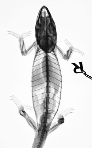
Fig. 1. Dorsoventral radiograph of the chameleon.
Plate VI
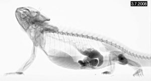
Fig. 2. Laterolateral contrast radiograph with barium sulphate.
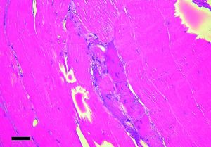
Fig. 3. Histopathology of the chameleon tongue showing scar of muscle fibres. HE × 10. Bar 50 μm.
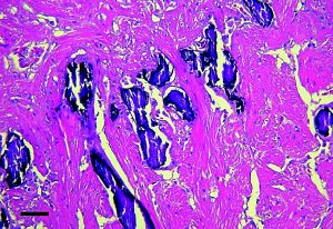
Fig. 4. The chameleon myocardium with a ‘layer’ of mineralization foci. HE × 10. Bar 50 μm.
Plate VII
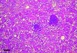
Fig. 5. Several small bacterial abscesses in the chameleon liver. HE × 10. Bar 50 μm.
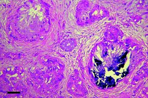
Fig. 6. The chameleon kidneys. Uric acid tophi with mineralization. HE × 10. Bar 50 μm.